


 Language
 Clinical Specialties
Dyspnoea, chest pain, unexplained deterioration – in acute medicine, every minute counts. Lung ultrasound provides bedside, radiation-free, real-time answers to the critical questions: Is there a pneumothorax? A pleural effusion? Cardiogenic pulmonary edema? This article shows what each finding looks like – and what it means clinically.
Key Takeaways
- A-lines are horizontal reverberation artifacts indicating a normally aerated lung.
- B-lines arise when fluid is retained in lung tissue – three or more per acoustic window indicate pulmonary congestion or interstitial syndrome.
- Absent lung sliding is the cardinal sign of pneumothorax – the accumulation of air in the thoracic cavity.
- Pleural effusion appears as echo-free fluid between the pleural layers and is detectable by sonography with greater sensitivity than on plain radiography.
- Handheld ultrasound such as the Vscan Air enables lung ultrasound directly at the bedside – without a cart, without time lost.
What Is Lung Ultrasound? Definition and Clinical Principles
Lung ultrasound – also known as lung sonography or thoracic sonography – is a point-of-care procedure for bedside assessment of the lungs, pleura, and adjacent structures. Unlike conventional organs, the lung does not produce a direct echo structure: air reflects ultrasound waves completely.
What lung ultrasound uses instead are characteristic imaging patterns – so-called artifacts, i.e., signals that do not directly depict an organ but arise from the interaction of sound and pleura, and allow reliable conclusions about the state of the lung tissue.
The procedure is radiation-free, repeatable, and can be performed with the patient supine or sitting. Meta-analyses show that lung ultrasound has higher sensitivity with comparable specificity compared to conventional chest X-ray for pleural effusion, pneumonia, pneumothorax, and pulmonary edema. (Staub et al., Radiology: Cardiothoracic Imaging, 2021)
Why Is Lung Ultrasound Often Underestimated?
For a long time, the lung was considered an “ultrasound-hostile” organ – too much air, too little signal. This assumption has proven false. Precisely because air completely reflects sound, characteristic artifacts arise that are clinically highly relevant. Clinicians who can distinguish A-lines from B-lines and assess lung sliding can make well-founded decisions – without radiation, without transport.
Another reason for underestimation: lung ultrasound was considered difficult to learn. Current studies and AI-assisted tools show, however, that reliable findings are achievable even with limited experience – particularly when structured protocols and image guidance are used.
When Is Thoracic Ultrasound Clinically Indicated?
Lung ultrasound is not a substitute for comprehensive diagnostics – but it is a powerful first tool when rapid answers to targeted questions are needed.
Acute Dyspnoea and Breathlessness
Dyspnoea is one of the most common emergency presentations. Possible causes include cardiogenic pulmonary edema, pneumonia, pleural effusion, or pneumothorax. Acute exacerbation of COPD or bronchial asthma is also possible. Lung ultrasound enables rapid differentiation: bilateral B-lines suggest congestion, absent lung sliding suggests pneumothorax, echo-free fluid suggests effusion, and subpleural consolidations suggest pneumonia.
What is often overlooked: the examination is also gentler for the patient. Karen Kelly, Advanced Nurse Practitioner and founder of HeartPath in Dublin, puts it simply: “You don’t have to lie flat when you have difficulty breathing. The examination can be performed in a few seconds, even sitting up.”
Chest Pain
In acute chest pain, sonography substantially broadens the diagnostic spectrum. Pericardial effusion, pleural effusion, pneumothorax, and pleural consolidations are directly visualizable on ultrasound. Combined with cardiac POCUS for assessment of cardiac function and the pericardium, the range of life-threatening causes can be rapidly narrowed down.
Further Indications
The range of applications extends well beyond the emergency department. Whether monitoring a pleural effusion, tracking pneumonia, guiding thoracentesis, or checking after chest drain insertion – lung ultrasound is clinically established in all these situations. It is gaining particular relevance in general practice, where no X-ray equipment is available. More on this is shown in the webinar on dyspnoea and chest pain with AI-assisted handheld ultrasound.
Fundamentals of Lung Ultrasound
Three artifacts and one phenomenon form the foundation of lung sonography. Clinicians who can reliably recognize them can answer most clinically relevant questions at the bedside.
A-Lines – Signs of Normal Lung
A-lines are horizontal, bright reverberation artifacts that appear at equal intervals below the pleural line. They arise from reflection of the ultrasound beam off the air-filled lung and are normal – they indicate normal aeration at the site examined. Where A-lines predominate and lung sliding is present, pneumothorax at that site is excluded.
B-Lines – Identifying Interstitial Syndrome
B-lines are vertical, bright, laser-like signals that extend from the pleural line to the bottom of the image and move with the respiratory cycle. They arise when lung tissue is thickened by retained fluid – in pulmonary edema, pneumonia, lung contusion, or interstitial lung disease.
Three or more B-lines per acoustic window are considered pathological. Bilateral detection in at least two regions is associated with pulmonary congestion with a sensitivity of 100% and specificity of 92%. (Volpicelli et al., Intensive Care Med, 2012) Important: B-lines exclude pneumothorax at the examined site – as they indicate that the lung is still in contact with the chest wall there.
Lung Sliding
Lung sliding refers to the visible movement of the two pleural layers against each other during the respiratory cycle – a normal sign of ventilation. In M-mode, it appears as the seashore sign (sandy pattern). Absent lung sliding is the most important sign of pneumothorax. If the lung point is additionally detectable, the diagnosis is considered confirmed.
The sensitivity of lung ultrasound for pneumothorax is 87–99%, and specificity 98–99% – significantly higher than supine chest X-ray. (Ding et al., Chest, 2011)
Key Pathological Findings in Lung Ultrasound
The three most common pathological findings in lung ultrasound – pleural effusion, pneumothorax, and consolidation – are reliably detectable sonographically and can be directly applied clinically.
Pleural Effusion: Classification and Clinical Relevance
A pleural effusion – a fluid collection between the two pleural layers – appears as a dark, echo-free zone on ultrasound. Typical finding: an echo-free zone between the lung and diaphragm on lateral or posterior oblique imaging. Sonographically, even small effusions of approximately 20 ml can be detected – on chest X-ray, effusions are reliably visible only from approximately 200–300 ml (Sikora et al., International Scholarly Research Notices, 2012).
Effusion volume can be estimated from the maximum effusion height in the supine position: 1 cm corresponds roughly to 200 ml, 2 cm to approximately 500 ml (Balik et al., Intensive Care Medicine, 2006). Inflammatory and non-inflammatory fluid cannot be reliably distinguished sonographically – however, effusions with visible internal echoes suggest inflammation, pus, or blood.
Detecting and Excluding Pneumothorax
A pneumothorax occurs when air enters between the two pleural layers, compressing the lung. The most important sonographic sign is absent lung sliding – the absence of the normal gliding motion of the pleural layers during breathing. Further indicators include absent B-lines and no lung pulse. If the lung point is detectable, the diagnosis is confirmed.
For exclusion: as soon as lung sliding, B-lines, or a lung pulse – the vibration of the lung transmitted by the heartbeat – are detectable, pneumothorax at the examined site can be excluded with very high probability. (Ding et al., Chest, 2011)
Consolidation: Pneumonia or Atelectasis?
Consolidation appears on ultrasound as a dark, tissue-like area beneath the pleural line – also called the “hepatization sign”. Two common causes underlie this finding: pneumonia, where inflammation densifies lung tissue, or atelectasis, where parts of the lung collapse and are no longer aerated.
The difference is seen in the air bronchograms – bright air signals within the airways inside the consolidation. If they move with the respiratory cycle, this suggests pneumonia. If they are static, this more likely indicates atelectasis.
In pneumonia, a concomitant pleural effusion can frequently be detected sonographically – in 40–60% of patients with bacterial pneumonia. (StatPearls, NCBI, 2023)
Echocardiography as a Complement in Acute Thoracic Ultrasound
Lung and heart can be examined in a single session – on the same device, without interruption. Particularly in dyspnoea, the combination of lung ultrasound and cardiac POCUS is the diagnostic standard.
Cardiac POCUS in Dyspnoea
The question “Is the dyspnoea of cardiac or pulmonary origin?” can be answered in a targeted manner at the bedside. Impaired cardiac function combined with bilateral B-lines suggests cardiogenic pulmonary edema. Normal cardiac function with unilateral B-lines or consolidation directs suspicion towards pneumonia or effusion.
Dr Guy Lloyd, Head of Echocardiography at Bart’s Heart Center in London, summarizes the clinical potential: “I can make 90% of the diagnoses I need right at the bedside and treat the patient immediately.”
Pericardial Effusion and Signs of Tamponade
Pericardial effusion appears as a dark fluid rim between the myocardium and pericardium. In tamponade – when the effusion grows large enough to compress the heart externally and impair filling – signs of right ventricular diastolic collapse are visible on ultrasound. The probe can be placed subxiphoidally or laterally to the sternum – usually sufficient for rapid assessment even in poor acoustic conditions.
Lung Ultrasound with Mobile Ultrasound – What AI-Assisted Handheld Devices Offer
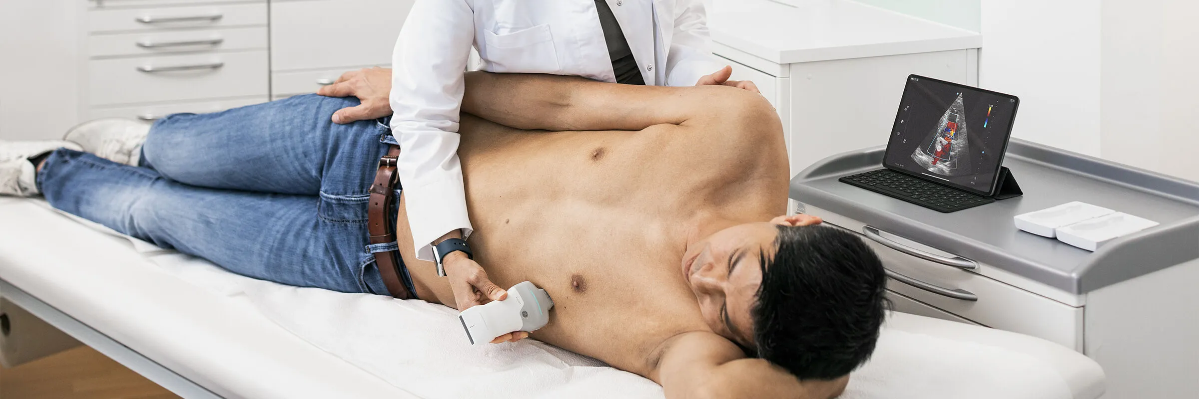
Lung ultrasound benefits particularly from the mobility of wireless handheld devices. What once required a wheeled ultrasound cart now works with a device in the coat pocket – in general practice, the ambulance, or directly at the bedside in intensive care.
Advantages of Handheld Ultrasound in Lung Sonography
Wireless ultrasound devices for general medicine such as the Vscan Air CL combine curved-array and linear probes in a single device. The curved-array probe is suitable for assessing pleural effusion, interstitial syndrome, and consolidations. The linear probe offers higher resolution for near-field assessment of the pleura – ideal for pneumothorax diagnostics and subpleural lesions.
Vscan Air SL + Caption AI – AI Support for Heart & Lung
The Vscan Air SL with Caption AI supports not only automatic EF measurement (cardiac pumping function), but also structured image acquisition in combined cardiac and pulmonary examinations. Caption Guidance™ guides the examiner through standardised imaging planes, which is particularly valuable for users who perform cardiac and pulmonary POCUS infrequently. More on the clinical application is shown in the webinar on dyspnoea and chest pain with AI-assisted handheld ultrasound.
Frequently Asked Questions about Lung Ultrasound
Can Lung Ultrasound Replace a Chest X-Ray?
Not entirely – but in many emergency situations it is superior. For pneumothorax, pleural effusion, pulmonary edema, and pneumonia, lung ultrasound shows higher sensitivity than chest X-ray in meta-analyses. It is radiation-free, immediately available, and performable at the bedside. For assessment of deep lung tissue, the mediastinum – the area between the lungs containing the heart and great vessels – or bony structures, plain radiography or CT remains superior.
How Can I Reliably Exclude Pneumothorax by Ultrasound?
Pneumothorax can be practically excluded at the examined site if lung sliding, B-lines, or a lung pulse are detectable. The examination should be performed bilaterally and systematically – preferably along the mid-clavicular line and the lateral chest wall.
How Do I Identify a Pleural Effusion – and How Do I Estimate the Volume?
A pleural effusion appears as an echo-free zone between the lung and diaphragm on lateral or dorsal oblique imaging. Volume can be estimated from the maximum effusion height in the supine position: a height of 1 cm corresponds roughly to 200 ml, 2 cm to approximately 500 ml (Balik et al., Intensive Care Medicine, 2006). For more precise calculations, validated formulae are available, such as the Balik formula.
How Many B-Lines Are Clinically Relevant for Interstitial Syndrome?
From three B-lines per acoustic window, the finding is considered pathological. Interstitial syndrome – fluid retention in lung tissue indicating various conditions such as pulmonary edema or pneumonia – is diagnosed when this finding is present bilaterally in at least two regions. One to two B-lines can be normal – particularly in the lower lung zones.
How Long Does a Lung Ultrasound Examination Take?
A focused lung ultrasound with four to eight acoustic windows takes 3–5 minutes in experienced hands. A complete thoracic sonography including cardiac POCUS can be performed in 10–15 minutes. Regular training and a device that is always ready to use are prerequisites.
Which Probes Are Suitable for Lung Ultrasound?
For most indications – pleural effusion, interstitial syndrome, consolidations – the curved-array probe (2–5 MHz) is appropriate. For assessment of the pleura directly beneath the skin surface – pneumothorax, superficial lesions – the linear probe (5–12 MHz) is the better choice. Both are available on a single device with the Vscan Air CL.
JB09745XE
Other Topics

How handheld ultrasound can impact practice in Primary Care – featuring GE HealthCare's Vscan Air™ family with Vscan Air™ CL and Vscan Air™ SL

Mobile Ultrasound in Emergency Medical Services: Use, Benefits, and Selection

Ejection Fraction: Significance, Normal Values, and Measurement

Bladder Volume Measurement by Ultrasound: Methods, Formula, and Application

MSK Medicine: Musculoskeletal Ultrasound in Clinical Practice

Empowering nurses through innovation: Vscan Air™ handheld ultrasound in action at the ICN Congress 2025

A better outlook for mothers in Ethiopia: How Vscan Air™ is transforming maternal care in remote regions

The benefits of handheld ultrasound with AI-based Auto Bladder Volume measurement in everyday clinical practice

Experiences with handheld ultrasound in obstetrics

Vscan Air™ CL ultrasound combined with Usono ProbeFix Dynamic: Observation of muscle activity in real-time



 Belgium
  Brasil
  Bulgaria
  Croatia
  Czechia
  Denmark
  Deutschland
  España
  Estonia
  Finland
  France
  Greece
  Hungary
  Ireland
  Italia
  Latvia
  Lithuania
  Luxembourg
  Malaysia
  Netherlands
  Norway
  Österreich
  Poland
  Portugal
  Romania
  Slovakia
  Slovenia
  Sweden
  Schweiz
  UK
UK 
