


 Language
 Clinical Specialties
Sonographic bladder volume measurement is an established procedure for non-invasive assessment of the urinary bladder. In clinical practice, it is used in particular to evaluate bladder filling, post-void residual, and possible urinary retention. The method is fast, radiation-free, and can be performed directly at the bedside.
Key Takeaways
- Ultrasound bladder volume measurement determines the filling volume of the urinary bladder in real time.
- It is used to assess post-void residual, urinary retention, and bladder emptying disorders.
- The examination is non-invasive, radiation-free, and suitable as a point-of-care procedure.
- Volume is calculated manually or automatically via integrated software solutions.
- Modern systems can increase reproducibility and reduce operator-dependent measurement errors.
What Is Ultrasound Bladder Volume Measurement?
Ultrasound bladder volume measurement is a non-invasive imaging procedure for determining the filling status of the urinary bladder. It is based on sonographic visualization of the bladder in multiple planes, followed by volume calculation.
In everyday clinical practice, it is used primarily to assess post-void residual after voiding (urination) and to investigate possible urinary retention. It is part of basic urological diagnostics and is also routinely used in geriatrics and general medicine.
A key advantage lies in the immediate availability of the examination at the bedside. This allows findings to be incorporated promptly into further diagnostic and therapeutic decision-making.
The method can be performed manually using geometric approximation formulae or automatically through integrated software solutions. Automated procedures can help to improve comparability of measurement results and reduce dependency on examiner experience.
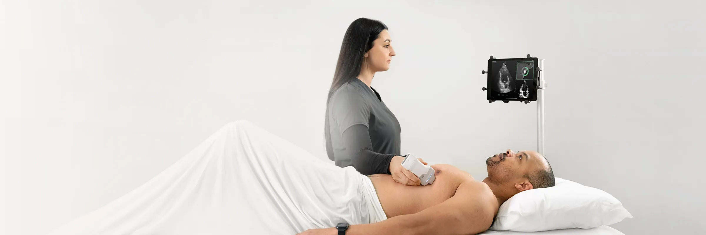
In the context of modern point-of-care ultrasound (POCUS) in medicine, mobile bladder sonography is therefore gaining increasing significance. As Dr Martin Altersberger, a cardiologist from Steyr, confirms: “It should be used as early as possible to detect serious conditions. We want to identify patients with relevant conditions, whether young or old, for whom we can make a difference and have a positive impact through early diagnosis and subsequent treatment.”
When Sonographic Bladder Volume Measurement Is Clinically Relevant
Ultrasound bladder volume measurement is used when the filling status of the urinary bladder or a possible post-void residual needs to be clinically assessed. It provides timely information for clinical interpretation without requiring invasive measures such as catheterization (Resnick et al., Rehabilitation Nursing, 1995).
Indications in Urology and General Medicine
In urology, bladder volume measurement is among the established procedures used when urinary retention or bladder emptying disorders are suspected. It is also used for follow-up in prostatic hyperplasia or after surgical procedures.
In general medicine, the examination plays an important role in non-specific symptoms such as lower abdominal pain, voiding problems, or incontinence. It enables a rapid initial assessment without invasive measures. Particularly in the GP setting, an ultrasound device for general medicine supports rapid assessment directly at the bedside or in the consulting room.
In inpatient geriatrics the method is also particularly relevant, as many patients cannot reliably report symptoms. Studies on ultrasound bladder volume measurement in geriatrics show that the method supports objective assessment of bladder function in everyday nursing care and facilitates clinical decision-making.
Considerations in Children and Urogynecology
In children, bladder volume measurement is used to investigate functional bladder emptying disorders. It is particularly relevant in enuresis or suspected post-void residual.
In urogynecology, it serves to assess postpartum changes or functional disorders of the pelvic floor. The examination is likewise non-invasive and can be performed repeatedly.
Post-Void Residual Measurement in the Clinical Decision Process
Interpretation of bladder volume and post-void residual measurements depends substantially on the overall clinical situation and should always be made in the context of symptoms, history, and physical examination.
The timing of measurement after voiding also influences the informative value. A measurement taken too early can overestimate post-void residual because bladder emptying is not yet complete. Standardized measurement timing therefore improves comparability of results in everyday clinical practice.
In cases of unclear or borderline findings, follow-up measurements are useful to distinguish measurement variability from a genuine functional disorder. The sonographic measurement provides objective volume data for assessing bladder function and supports clinical decision-making within the overall context of the patient’s situation.
Ultrasound measurement allows rapid assessment after voiding and can in many cases reduce the need for catheterization. Studies describe good agreement between sonographic post-void residual measurement and catheter measurement (Manfredini et al., French Journal of Urology, 2025). An ultrasound device for nursing care with a mobile solution, such as the Vscan Air CL, can additionally facilitate standardized post-void residual checks directly at the bedside.
Post-void residual measurement is therefore a central component of the urological and internal medicine decision process, linking diagnostic imaging directly to clinical action.
Clinical Interpretation and Measurement Quality in Daily Practice
Interpretation of bladder volume and post-void residual measurements depends substantially on the overall clinical situation. Individual values should therefore always be assessed in the context of symptoms, history, and physical findings.
The timing of measurement after voiding also influences the informative value. If measurement is taken too early, post-void residual can be overestimated because bladder emptying is not yet fully complete. A standardized procedure with defined measurement timing therefore improves the comparability of results in everyday clinical practice.
In cases of unclear or borderline findings, repeat measurements are useful. They help to distinguish measurement variability from a true functional bladder emptying disorder.
Sonographic measurement provides objective volume data for assessing bladder function. It supports clinical decision-making and improves assessment within the overall context of the patient’s situation.
How to Measure Bladder Volume with Ultrasound: Step by Step
Sonographic bladder volume measurement is performed in a few standardized steps. The aim is a reproducible and largely artifact-free volume determination in everyday clinical practice.
Preparation, Probe, and Device Settings
The patient ideally lies supine with a relaxed abdominal wall. A convex or sector probe is placed suprapubically. The image depth is chosen so that the urinary bladder is fully captured.
Measurement in Sagittal and Transverse Planes
First, the bladder is measured in the sagittal plane, then in the transverse plane. Length, width, and height of the bladder are recorded.
The values are then entered into the volume formula or calculated automatically. Correct plane selection is critical for measurement accuracy.
The Formula for Calculating Bladder Volume
Ultrasound bladder volume measurement is based on a geometric approximation formula. It enables a rapid estimate of bladder volume from three spatial dimensions.
Standard Formula and Correction Factor 0.5 to 0.7
The standard formula is: Volume ≈ length × width × height × 0.5–0.7.
The correction factor accounts for the individual shape of the urinary bladder, which is rarely exactly spherical or ellipsoidal.
Depending on the study and device, a factor between 0.5 and 0.7 is used. The choice of factor influences the accuracy of the volume estimate.
The calculation can be performed manually or automatically by integrated software. Modern systems thereby reduce both computational and operator-dependent sources of error.
Calculation in Children
The same basic formula is used in children but adapted to smaller volumes. Here, accuracy depends particularly on precise plane positioning.
Reference values are age-dependent and must be interpreted clinically. Interpretation in the context of symptoms and drinking habits is decisive.
Normal Values and Pathological Findings
Assessment of bladder volume depends on filling status, age, and clinical situation. Correct interpretation in the context of symptoms and post-void residual is decisive.
In adults, a post-void residual of less than 50 ml is generally considered unremarkable. Values above 100–150 ml may indicate impaired bladder emptying. Higher volumes are clinically relevant and should be investigated further. This classification is also consistent with urological guidelines on the diagnosis of bladder emptying disorders, which describe ultrasound measurement of post-void residual as the standard procedure.
In children, reference values are age-dependent and considerably more variable. Here the overall clinical situation – not a single threshold value – is what matters.
Sources of Error and Accuracy of Ultrasound Measurement
The accuracy of bladder volume measurement depends on several factors. Correct visualization of the bladder, choice of imaging planes, and the user’s experience are decisive.
Common errors arise from incomplete visualization of the bladder or an asymmetric shape that is not correctly captured. Bladder filling that is too high or too low can also affect measurement accuracy.
Device settings also play an important role. Incorrect depth settings or an unsuitable probe can compromise image quality and therefore volume calculation. Automated systems can reduce this variability by algorithmically detecting the bladder contour and performing standardized calculations.
Everyday clinical practice makes clear how profoundly point-of-care ultrasound is changing decision-making. As Paul Stoneman, Advanced Nurse Practitioner at Beaumont Hospital in Dublin, describes: “When I’m faced with a cardiac emergency, I don’t reach for the stethoscope – I reach for the Vscan. In moments like that, the handheld ultrasound gives me real-time information that guides my decisions.”
Manual Formula Calculation Compared with AI-Assisted Measurement
Manual volume calculation depends on the correct measurement of the three spatial axes. This introduces a degree of operator dependency in everyday clinical practice.
AI-assisted systems can support the standardization of volume measurement and thereby potentially contribute to reducing measurement variability.
Bladder Volume Measurement with the Vscan Air CL with Auto Bladder Volume
Sonographic bladder volume measurement is increasingly part of a mobile point-of-care workflow. Digital systems support rapid, standardized volume determination directly at the patient’s side.
The Vscan Air CL with Auto Bladder Volume enables flexible use in hospitals, practices, and nursing care. The wireless design facilitates use in everyday clinical practice and at the bedside.
With the Auto Bladder Volume function, the Vscan Air CL can calculate bladder volume automatically. This reduces operator dependency and improves measurement reproducibility.
Integration into existing workflows supports rapid clinical decisions. This is particularly relevant in urology, geriatrics, and emergency medicine.
Frequently Asked Questions about Ultrasound Bladder Volume Measurement
Which Bladder Volume Is Considered Normal?
In sonographic assessment, post-void residual after voiding (micturition) is generally evaluated. In adults, a post-void residual of less than 50 ml is usually considered unremarkable.
Values between 50 and 100 ml are borderline and should be monitored in the clinical course. From approximately 100–150 ml, a clinically relevant post-void residual with possible voiding disorder is commonly diagnosed.
How Is Bladder Volume Calculated in Children?
In children, the same basic formula is used as in adults. However, interpretation is age-dependent, as reference values vary considerably. Clinical assessment in combination with symptoms such as enuresis (bedwetting) or voiding disorders is therefore paramount.
Which Probe Is Suitable for Bladder Sonography?
A convex probe is generally used for bladder sonography. It provides sufficient penetration depth and complete visualization of the urinary bladder. In children or slim patients, a linear probe may suffice if the bladder lies superficially.
How Accurate Is Sonographic Measurement Compared with Catheterization?
Ultrasound measurement generally provides a good clinical approximation of the true bladder volume. The diagnostic accuracy of bladder scan measurement has been described in clinical studies as sufficient for routine diagnostics, particularly with standardized use. Catheterization remains the reference standard in individual cases, but is invasive and unsuitable for routine diagnostics.
JB09714xe
Other Topics

How handheld ultrasound can impact practice in Primary Care – featuring GE HealthCare's Vscan Air™ family with Vscan Air™ CL and Vscan Air™ SL

Lung Ultrasound: Recognizing Findings and Making Diagnoses

Mobile Ultrasound in Emergency Medical Services: Use, Benefits, and Selection
Ejection Fraction: Significance, Normal Values, and Measurement

MSK Medicine: Musculoskeletal Ultrasound in Clinical Practice

Empowering nurses through innovation: Vscan Air™ handheld ultrasound in action at the ICN Congress 2025

A better outlook for mothers in Ethiopia: How Vscan Air™ is transforming maternal care in remote regions

The benefits of handheld ultrasound with AI-based Auto Bladder Volume measurement in everyday clinical practice

Experiences with handheld ultrasound in obstetrics

Vscan Air™ CL ultrasound combined with Usono ProbeFix Dynamic: Observation of muscle activity in real-time



 Belgium
  Brasil
  Bulgaria
  Croatia
  Czechia
  Denmark
  Deutschland
  España
  Estonia
  Finland
  France
  Greece
  Hungary
  Ireland
  Italia
  Latvia
  Lithuania
  Luxembourg
  Malaysia
  Netherlands
  Norway
  Österreich
  Poland
  Portugal
  Romania
  Slovakia
  Slovenia
  Sweden
  Schweiz
  UK
UK 
