


 Language
 Clinical Specialties
A mobile ultrasound device is now standard equipment on modern emergency response vehicles — from physician-staffed emergency response vehicles to medical helicopters. It delivers findings at the scene that used to be available only at the hospital. This article shows where mobile ultrasound delivers the greatest benefit in emergency medicine, which protocols are well established, and what matters most when selecting a device.
Key Takeaways
- Mobile ultrasound devices enable life-saving diagnoses directly at the scene — without losing time to transport.
- FAST and E-FAST are the most important emergency protocols for trauma patients and can be performed in the prehospital setting in under 3 minutes.
- According to a position statement from the European Association of Echocardiography, experienced users can perform a valid focused cardiac assessment with handheld devices in about 4 minutes.
- Image quality and ruggedness are the decisive selection criteria for EMS use — alongside IP67 certification and MIL-STD-810G drop protection.
- Vscan Air CL and SL are CE-certified medical devices for prehospital and clinical use — from abdominal trauma to initial cardiac care.
What is a mobile ultrasound device? Definition and scope
A mobile ultrasound device is a compact ultrasound system that can be operated without fixed infrastructure — at the scene, in an ambulance, in a helicopter, or directly at the patient's bedside. Unlike conventional console systems, it does not require a permanent location and runs independently of mains power.
Differences between handheld, laptop, and cart-based ultrasound
Ultrasound devices relevant to EMS can be grouped into three broad device classes — each with different strengths depending on the use case.
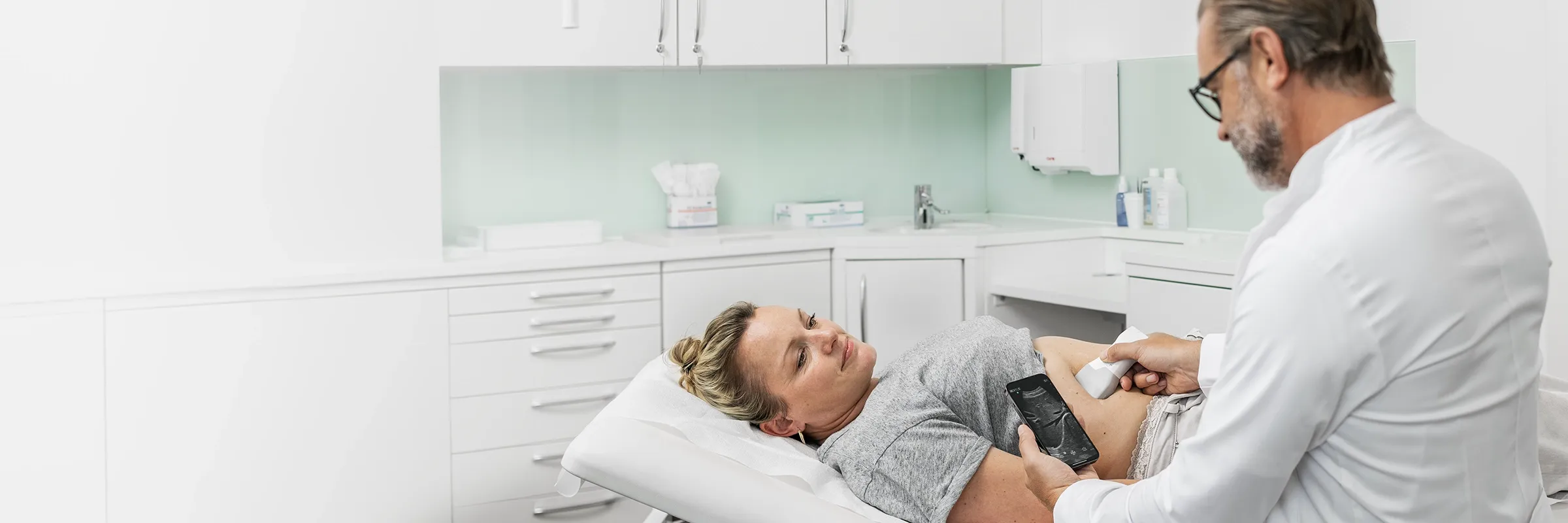
Handheld devices are the most compact form factor and the best fit for prehospital use. A wireless probe connects via an app to a smartphone or tablet. Modern devices typically weigh under 300 g and are ready to use within seconds.
Laptop-based devices are compact, portable systems with their own display and battery — heavier than handheld devices, typically between 2 and 5 kg. They usually offer more imaging modes than handheld devices. They are less suitable for EMS use due to their weight and limited ruggedness.
Cart-based systems are stationary console devices designed for the hospital setting. They deliver the highest image quality and the broadest feature set but are not designed for prehospital use.
Features of a modern mobile ultrasound device
A modern handheld device for EMS use should meet the following criteria:
- Wireless connectivity via Wi-Fi or Bluetooth — no cables to get in the way during a call
- Ruggedness to MIL-STD-810G (military drop test standard) and IP67 certification (waterproof and dustproof)
- Dual-probe design for different applications without switching devices
- Battery life of at least 45–60 minutes to cover a full call
- DICOM interface for direct data transfer to the hospital system on arrival
- Intuitive touchscreen operation — even while wearing gloves
Why mobile ultrasound is critical in emergency medicine
In emergency medicine, minutes matter. Diagnostic delays caused by transport, wait times, or the absence of imaging at the scene eat into the time available for treatment. Mobile ultrasound shortens that window substantially — and improves the basis for clinical decisions at the same time.
Diagnostic confidence and time savings at the scene
Prehospital ultrasound diagnostics make it possible to identify life-threatening findings before the patient even arrives at the hospital. That shapes the choice of destination facility, the advance notification of the trauma team, and how transport is prioritized. An early finding also cuts down on unnecessary additional workup in the ED and speeds up handoff.
For the FAST protocol in trauma patients, a systematic review and meta-analysis of 1,356 patients reports a pooled specificity of 0.97 for the detection of free intra-abdominal fluid, with a sensitivity of 0.60. (Lin et al., BMC Emergency Medicine, 2024) That means a positive finding is highly reliable. A negative finding does not rule out injury and must be interpreted in clinical context.
Focused cardiac assessment is also feasible in the prehospital setting. According to a position statement from the European Association of Echocardiography, experienced users can perform a valid bedside evaluation of cardiac structures, the pleural space, and the great abdominal vessels with handheld devices in about 4 minutes. (Sicari et al., European Journal of Echocardiography, 2011)
Shorter exam times in everyday clinical practice
In-hospital, mobile ultrasound shortens the diagnostic workup just as significantly. The BLUE protocol — a structured exam scheme for quickly identifying the cause of acute respiratory distress — can be completed in under 3 minutes and differentiates the most common causes of acute respiratory failure. (Lichtenstein & Mézière, Chest, 2008)
Regular use also builds operator competence. Studies show that frequent use of point-of-care ultrasound (POCUS) measurably increases diagnostic confidence and speed. (El Sayed et al., Journal of Emergencies, Trauma, and Shock, 2018) In the ED, bedside ultrasound often replaces time-consuming X-ray or CT scans for targeted clinical questions — with no radiation exposure and no need to move the patient.
Use cases in EMS and emergency medicine
Mobile ultrasound has wide-ranging applications in emergency medicine. The most important areas of use are trauma, dyspnea, cardiac emergencies, and vascular access. It is also seeing growing use in obstetrics, aesthetic medicine, and musculoskeletal medicine — for example, in bedside evaluation of tendons, ligaments, and joints in MSK ultrasound.
Ground EMS and air rescue
In ground EMS, the handheld device makes it possible to scan directly at the scene or inside the ambulance. Wireless devices that don't depend on a cart have a clear advantage here — they can be used even in cramped conditions.
Air rescue imposes its own requirements. Vibration, tight quarters, and often poor lighting call for intuitive devices with a bright display and simple one-handed operation. IP67 certification ensures the device stays operational in rain or contamination. Devices intended for use in rescue helicopters must meet the applicable aviation regulations.
Emergency department and rural care
In the ED, mobile ultrasound is a fixed part of the primary diagnostic workup. Wireless handheld devices make it possible to integrate ultrasound into the trauma team's workflow without rolling up a second console unit.
In rural regions with long transport times, prehospital diagnosis carries extra weight. Early identification of internal bleeding or a pericardial effusion — a fluid collection around the heart — can directly determine the choice of destination facility and, with it, the patient's chance of survival. A faster diagnosis can also shorten the wait and reduce uncertainty for patients.
For an overview of the clinical use of ultrasound devices in emergency medicine, see our specialty page.
Clinical applications: emergency ultrasound protocols
Emergency ultrasound follows structured protocols built around a small set of targeted clinical questions. They don't require full sonography training, but they do require solid initial instruction and regular practice.
FAST and E-FAST in the trauma patient
FAST ultrasound (Focused Assessment with Sonography for Trauma) is the most widely used emergency ultrasound protocol in the world. It scans four windows for free fluid — a sign of internal bleeding: the pericardium, the right upper quadrant (Morison's pouch), the left upper quadrant (Koller's pouch), and the pelvis. In experienced hands, the exam takes under 3 minutes.
Extended FAST (E-FAST) adds thoracic ultrasound to the classic protocol: bilateral detection or exclusion of a pneumothorax — an abnormal collection of air in the chest — and a hemothorax. According to a meta-analysis of 24,350 patients, the specificity of E-FAST for detecting a pneumothorax is 99% and the sensitivity is 69%. (Netherton et al., Canadian Journal of Emergency Medicine, 2019)
POCUS for dyspnea and chest pain
In acute respiratory distress or chest pain, Point-of-care ultrasound (POCUS) in medicine allows rapid differentiation of the most common causes. Bilateral B-lines point to pulmonary edema — fluid in the lungs; absent pleural sliding points to a pneumothorax; a dark fluid zone points to a pleural effusion.
The BLUE protocol structures this exam in a few steps and is established in ICUs and EDs worldwide. (Lichtenstein & Mézière, Chest, 2008)
Ultrasound-guided vascular access and regional anesthesia
Ultrasound substantially improves accuracy when placing central and peripheral venous catheters — especially in patients with difficult venous anatomy. Studies show a clear reduction in complications and failed punctures compared with the blind technique. (Brass et al., Cochrane Database, 2015)
In regional anesthesia — for instance, when performing nerve blocks for pain management at the scene — ultrasound is also an established tool. Direct visualization of the nerve and the needle improves precision, reduces the risk of vascular puncture, and shortens procedure time. (Abrahams et al., British Journal of Anaesthesia, 2009)
Training and qualifications: what emergency physicians should know
The use of ultrasound in EMS is tied to qualification requirements. In Germany, the DEGUM tiered concept governs training in emergency ultrasound.
Supplementary emergency-medicine training and DEGUM certification
The German Society for Ultrasound in Medicine (DEGUM) has offered a structured training curriculum in emergency ultrasound since 2011, with three qualification levels. Level I covers the fundamentals of ultrasound diagnostics in the emergency setting; Levels II and III expand competence for more complex clinical questions and for teaching. (DEGUM, tiered concept for emergency ultrasound, 2011)
In Germany, paramedics may currently use ultrasound only under physician supervision or on a physician's order. Independent performance and interpretation are reserved for licensed physicians with the corresponding qualification. In other European countries — such as Norway and the Netherlands — prehospital ultrasound use by paramedics is already more widespread and more formally regulated.
Selection criteria for a mobile ultrasound device in EMS
The requirements in EMS differ markedly from those in the hospital. Ruggedness, speed, and ease of use take priority.
Image quality and transducer technology
Image quality is the most important clinical criterion. Only a clear image yields valid findings under time pressure. Modern handheld devices like the Vscan Air achieve diagnostically usable image quality despite their compact form factor. (Perez-Sanchez et al., The Ultrasound Journal, 2024)
The dual-transducer concept is particularly valuable for EMS: a curved-array probe for deep structures such as the abdomen and pericardium, and a linear probe for superficial structures such as the pleura and vessels — both on a single device, with no transducer swap.
Ruggedness, water resistance, and certifications (IP rating, MIL-STD-810G)
An EMS call is not a controlled lab environment. Rain, dust, impacts, and tight spaces are part of the job. A suitable device must:
- IP67-certified: fully dustproof and waterproof to a depth of 1 m for 30 minutes — a prerequisite for disinfection and outdoor use
- MIL-STD-810G tested: passed drop tests to the U.S. military standard — protection against impact damage in the field
- Suitable for air rescue: devices used in helicopters must meet the applicable aviation requirements
Battery life and wireless connectivity
A battery life of at least 45–60 minutes is necessary to cover a full prehospital call. Fast charging via the Qi standard allows topping off between calls without dealing with cables.
Wireless connectivity via Wi-Fi or Bluetooth streams images in real time to a smartphone or tablet — and, once the patient reaches the hospital, sends them via DICOM straight to the image archive (PACS). That saves time at handoff and ensures findings are not lost.
Mobile ultrasound with Vscan Air: built for use outside the hospital
The Vscan Air from GE HealthCare is a wireless dual-probe handheld system available in two configurations, designed for use under demanding conditions. Both models are certified to IP67 and MIL-STD-810G. (GE HealthCare, Vscan Air technical specifications)
Vscan Air CL for abdominal and thoracic emergencies
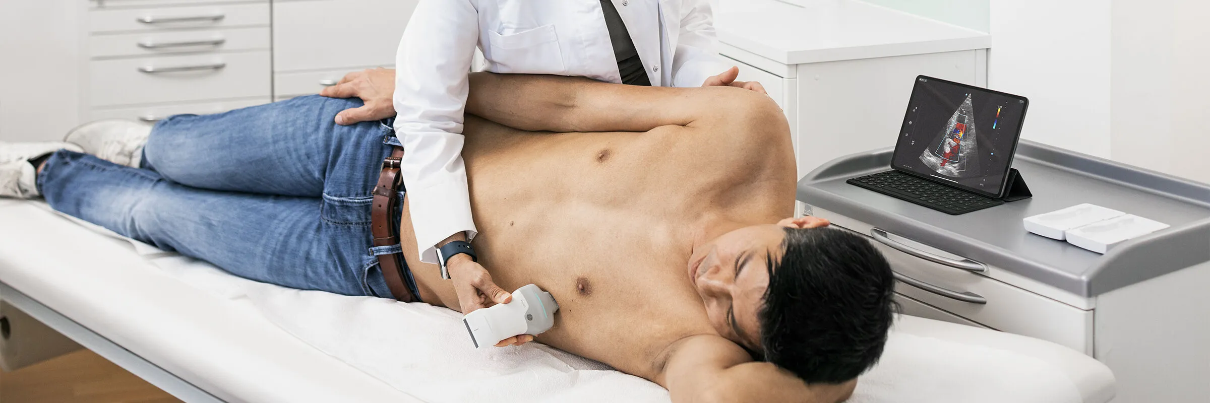
The Vscan Air CL combines a curved-array probe (2–5 MHz) with a linear probe (3–12 MHz). The curved-array probe is suited for FAST and E-FAST exams, assessment of the lungs and pleura, and imaging of deeper abdominal organs. The linear probe supports imaging of superficial structures, vascular access guidance, and pneumothorax diagnosis.
Vscan Air SL for cardiovascular first response
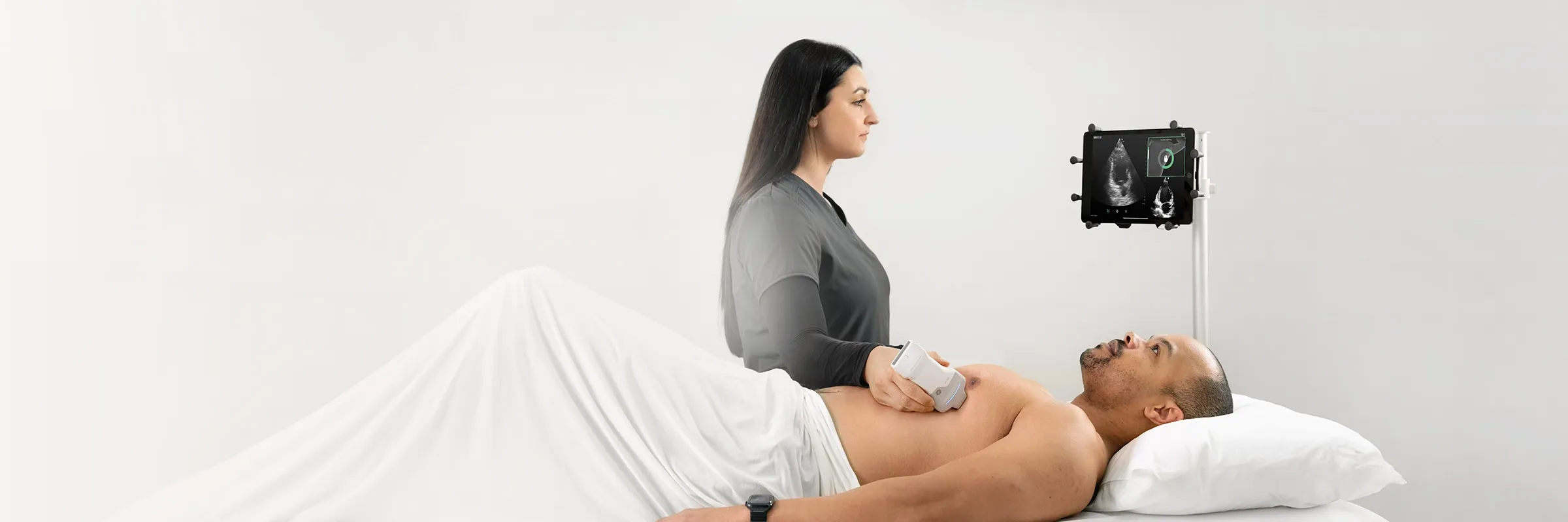
The Vscan Air SL combines a sector probe (1.6–3.7 MHz) with a linear probe. The sector probe is optimized for transthoracic cardiac ultrasound. Its compact footprint (22 × 17 mm) fits in narrow intercostal spaces and allows imaging of all four heart chambers. PW Doppler and M-mode are integrated as standard, enabling assessment of cardiac function, valve motion, and volume status.
Frequently asked questions about mobile ultrasound devices in EMS
How much does a mobile ultrasound device for EMS cost?
The price range is wide. Basic handheld devices start at roughly €2,000–3,000. Premium wireless dual-probe systems with clinically valid image quality, such as the Vscan Air, sit in the upper four-figure range. For EMS procurement decisions, we recommend reaching out directly to the manufacturer or an authorized reseller — including a review of financing options and service terms.
Are paramedics allowed to use a mobile ultrasound device?
In Germany, the independent performance and interpretation of ultrasound exams is reserved for licensed physicians with the appropriate qualification. Paramedics may use ultrasound devices under physician supervision or on a physician's order. The legal picture varies across Europe — in several countries, independent use by trained EMS personnel is already regulated and well established.
How rugged are wireless handheld ultrasound devices in the field?
Premium handheld devices like the Vscan Air are drop-tested to MIL-STD-810G and waterproof to IP67. That makes them suitable for use in EMS conditions — rain, dust, impacts. IP67 certification also allows routine disinfection, which is mandatory in EMS.
Which ultrasound transducer is best suited to EMS?
Probe choice depends on the primary use case. For FAST, E-FAST, and abdominal emergency diagnostics, the curved-array probe (2–5 MHz) is the first choice. For pneumothorax, vascular access, and superficial structures, the linear probe (5–12 MHz) is better suited. Devices with a dual-probe concept like the Vscan Air cover both application areas without a device swap.
References
- Sicari et al., European Journal of Echocardiography, 2011: The use of pocket-size imaging devices – a position statement of the European Association of Echocardiography. https://pubmed.ncbi.nlm.nih.gov/21810825/
- Lin et al., BMC Emergency Medicine, 2024: Prehospital ultrasound scanning for abdominal free fluid detection in trauma patients. https://doaj.org/article/ea3e29279b594003b1dd40da0b5f72a4
- Lichtenstein & Mézière, Chest, 2008: Relevance of lung ultrasound in the diagnosis of acute respiratory failure – the BLUE protocol. https://pubmed.ncbi.nlm.nih.gov/18403664/
- Netherton et al., Canadian Journal of Emergency Medicine, 2019: Diagnostic accuracy of E-FAST for pneumothorax. https://pubmed.ncbi.nlm.nih.gov/31317856/
- Brass et al., Cochrane Database of Systematic Reviews, 2015: Ultrasound guidance versus anatomical landmark-guided technique for central venous catheterisation. https://pubmed.ncbi.nlm.nih.gov/26408399/
- Abrahams et al., British Journal of Anaesthesia, 2009: Ultrasound guidance compared with electrical neurostimulation for peripheral nerve block – a systematic review and meta-analysis. https://pubmed.ncbi.nlm.nih.gov/19174373/
- Perez-Sanchez et al., The Ultrasound Journal, 2024: Comparison of 6 handheld ultrasound devices by point-of-care ultrasound experts. https://doaj.org/article/c9a6d041f150479abd9fcf43545441f3
- El Sayed et al., Journal of Emergencies, Trauma, and Shock, 2018: Prehospital ultrasound in trauma. https://pmc.ncbi.nlm.nih.gov/articles/PMC5852915/
JB09708xe
Other Topics

How handheld ultrasound can impact practice in Primary Care – featuring GE HealthCare's Vscan Air™ family with Vscan Air™ CL and Vscan Air™ SL
Lung Ultrasound: Recognizing Findings and Making Diagnoses
Ejection Fraction: Significance, Normal Values, and Measurement

Bladder Volume Measurement by Ultrasound: Methods, Formula, and Application

MSK Medicine: Musculoskeletal Ultrasound in Clinical Practice

Empowering nurses through innovation: Vscan Air™ handheld ultrasound in action at the ICN Congress 2025

A better outlook for mothers in Ethiopia: How Vscan Air™ is transforming maternal care in remote regions

The benefits of handheld ultrasound with AI-based Auto Bladder Volume measurement in everyday clinical practice

Experiences with handheld ultrasound in obstetrics

Vscan Air™ CL ultrasound combined with Usono ProbeFix Dynamic: Observation of muscle activity in real-time



 Belgium
  Brasil
  Bulgaria
  Croatia
  Czechia
  Denmark
  Deutschland
  España
  Estonia
  Finland
  France
  Greece
  Hungary
  Ireland
  Italia
  Latvia
  Lithuania
  Luxembourg
  Malaysia
  Netherlands
  Norway
  Österreich
  Poland
  Portugal
  Romania
  Slovakia
  Slovenia
  Sweden
  Schweiz
  UK
UK 
