


 Language
 Clinical Specialties
Musculoskeletal sonography has established itself as an integral part of diagnostics in orthopedics, rheumatology, and sports medicine. No other imaging modality combines rapid, dynamic, and radiation-free examination directly at the patient with real-time clinical correlation in this way. At the same time, mobile systems are increasingly making MSK ultrasound a point-of-care tool – from the outpatient setting to acute care.
Key Takeaways
The most important facts about MSK medicine and musculoskeletal ultrasound at a glance.
- MSK ultrasound examines muscles, tendons, ligaments, joints, and superficial nerves in real time.
- Unlike X-ray or CT, sonography is radiation-free and allows dynamic examinations under movement.
- Typical applications include the rotator cuff, Achilles tendon, carpal tunnel syndrome, joint effusions, and rheumatological inflammation.
- For superficial structures, high-frequency linear probes between approximately 5 and 15 MHz are used.
- Mobile systems such as the Vscan Air CL enable MSK examinations directly at the point of care without cables or carts.
What Is MSK Medicine? An Overview of the Fundamentals
MSK medicine (musculoskeletal medicine) focuses on the diagnosis and treatment of the locomotor system. This includes muscles, tendons, ligaments, joints, and bones. The term “MSK” stands for “musculoskeletal” and describes all structures responsible for posture and movement of the body.
MSK medicine brings together specialties such as orthopedics, rheumatology, sports medicine, trauma surgery, and physical medicine. The focus is on functional complaints, which are assessed clinically and with the help of imaging modalities.
This involves not only individual anatomical structures but also the assessment of complaints in the context of movement, loading, and function. The aim is to identify pain, functional disorders, and structural changes early and treat them in a targeted manner.
MSK Ultrasound as a Central Imaging Modality in Clinical Practice
MSK ultrasound has established itself as an important tool in modern MSK diagnostics. Unlike static modalities, it enables real-time examination under movement.
Sonography is particularly well-suited to structures of the active and passive locomotor system that lie superficially and can be displayed dynamically. These include muscles and tendons, as well as ligaments, joints, cartilage, and bursae. Typical findings in this context are:
- Tendon injuries or changes
- Muscle injuries
- Inflammation of the bursae (bursitis)
- Fluid in the joint (joint effusions)
- Superficial ligament injuries
- Inflammatory changes in rheumatic diseases
This makes functional problems visible directly during the examination – for example in shoulder impingement syndromes or tendon movements at the wrist. In point-of-care ultrasound (POCUS) the method is also gaining increasing importance, as examinations can be performed directly at the patient and without delay.
Evidence and Guidelines for MSK Sonography
The advantages of dynamic ultrasound diagnostics are clearly described by international professional societies, such as the European Federation of Societies for Ultrasound in Medicine and Biology (EFSUMB). The EFSUMB guidelines on MSK sonography emphasize in particular the importance of functional examination under movement and the high value of sonography in everyday clinical practice of musculoskeletal diagnostics (EFSUMB Guidelines and Recommendations for Musculoskeletal Ultrasound, Thieme 2022, Part 1 and Part 2).
These recommendations underline the value of dynamic assessment within musculoskeletal diagnostics, particularly for functional complaints and movement-dependent pathologies. In addition, evidence from systematic reviews shows that MSK ultrasound can achieve high diagnostic accuracy particularly for tendon and joint pathologies, especially in experienced hands (Dinnes et al., HTA 2003).
Further publications and reviews on musculoskeletal sonography can also be found in the GE HealthCare section for MSK publications and specialist literature.
MSK Ultrasound Compared with X-Ray, CT, and MRI
MSK ultrasound does not replace other imaging modalities but complements them effectively in many clinical situations.
X-ray is primarily suited to bony questions such as fractures or axis deviations. CT examinations provide high-detail resolution of complex bone structures but involve ionizing radiation. MRI, in turn, offers the most comprehensive visualization of deep soft-tissue structures but is more expensive and not always immediately available in everyday clinical practice.
MSK ultrasound plays to its strengths above all with superficial structures:
- Dynamic examination under movement
- Direct side-to-side comparisons
- Real-time visualization of tendons and muscles
- Radiation-free diagnostics
- Immediate availability at the point of care
Studies show that sonography can achieve high diagnostic value for certain questions – for example rotator cuff lesions or joint effusions – particularly in experienced hands. At the same time, MRI remains the reference standard for complex intra-articular or deep structures.
In clinical practice, the modalities frequently complement one another. Ultrasound is particularly suited to rapid initial assessment, follow-up, and dynamic examinations. MRI or CT is used in addition when deeper or more complex structures need to be assessed.
MSK Ultrasound: Method and Procedure
Musculoskeletal sonography is usually performed directly during the clinical examination. This allows complaints to be correlated immediately with the imaging findings.
A key advantage is that examinations do not have to be static. Patients can actively move joints or deliberately provoke specific pain points while the examination is ongoing.
In orthopedic or sports medicine consultations, this direct link between history, clinical examination, and imaging frequently shortens the diagnostic process considerably.
Probe Frequency, Imaging Planes, and Coupling
High-frequency linear probes are predominantly used for MSK examinations. Frequencies between approximately 5 and 15 MHz enable high resolution of superficial structures such as tendons, ligaments, or small joints.
Deeper regions – such as the hip – benefit instead from lower frequencies and wider acoustic fields.
The Vscan Air CL combines both approaches in a dual-probe system:
- Linear probe for superficial structures
- Curved-array probe for deeper anatomical regions
This eliminates the need to change probes during the examination. Particularly for time-critical or dynamic examinations, this can simplify the workflow.
Correct probe placement remains critical for image quality. Even small angular deviations can lead to artifacts such as anisotropy, particularly with tendons. Standardized imaging planes and practical experience are therefore among the most important fundamentals of MSK sonography.
Dynamic Examination of the Patient
Dynamic sonography is among the greatest advantages of MSK ultrasound.
While MRI and CT provide static image information, ultrasound allows assessment of movement patterns in real time. This makes functional complaints visible that often go unnoticed in resting examinations.
Typical examples include:
- Shoulder impingement during arm elevation
- Snapping tendons
- Tendon subluxations
- Nerve compressions during movement
- Instabilities of joint structures
In sports medicine and orthopedics, this dynamic approach frequently provides decisive additional information that is not visible on static modalities. In elite athletes, rapid assessment of acute complaints directly pitch side or in the training environment also plays an increasingly important role.
Typical Applications of MSK Ultrasound
Musculoskeletal ultrasound covers a broad spectrum of clinical questions – from acute sports injuries to diagnosis of chronic inflammation.
Shoulder, Elbow, and Wrist
The shoulder is among the most common applications of MSK sonography. The rotator cuff, subacromial bursa, and long head of the biceps tendon can be visualized in high resolution.
Examinations that incorporate movement are also very important here – for example, in so-called subacromial impingement. This assesses whether certain arm movements result in impingement in the shoulder region.
At the elbow, ultrasound is suitable for, among other things:
- Epicondylitis
- Biceps tendon pathologies
- Joint effusions
- Nerve irritations
At the wrist, carpal tunnel syndrome and tendon inflammation are among the most common indications. In addition, ganglia (benign fluid-filled protrusions at joints or tendons), tenosynovitis (inflammation of the tendon sheaths), and inflammatory changes in rheumatic diseases can frequently be assessed rapidly and without additional imaging.
Hip, Knee, and Ankle
At the hip, ultrasound can assess trochanteric bursitis (inflammation of the bursae in the lateral hip region), joint effusions, and tendon injuries, among other findings. As the hip lies deeper in the body than many other joints, curved-array probes are often used, allowing better visualization of deeper structures.
The knee is particularly well-suited to sonographic effusion diagnostics and the examination of superficial tendon and ligament structures.
At the ankle, the following questions are often paramount:
- Achilles tendon pathologies
- Ligament injuries
- Tendinopathies
- Joint effusions
Particularly in sports medicine, mobile sonography enables an immediate examination directly pitch side or in acute care. Decisions regarding further diagnostics or load-bearing capacity can thus be made more quickly.
Ultrasound-Guided Infiltrations
MSK ultrasound is used not only for diagnostics, but also to support targeted injections and minimally invasive treatments. Under ultrasound guidance, infiltrations can be performed more precisely, as the target structures remain directly visible during treatment. These include, among others:
- Joint injections
- Injections into the bursae (bursal infiltrations)
- Injections in the region of tendons
- Nerve blocks
Studies show that ultrasound-guided injections often achieve greater target accuracy than procedures without direct image control. At the same time, the real-time visualization of anatomical structures improves control during the intervention.
Qualification and DEGUM Certification for MSK Sonography
The quality of MSK ultrasound depends substantially on the examiner's experience. Structured training concepts that enable a step-by-step development of sonographic skills are therefore correspondingly important.
The German Society for Ultrasound in Medicine (DEGUM) has established a multi-tiered qualification system for musculoskeletal sonography in the area of the locomotor organs. This system ranges from basic courses through to expert certification. It also defines clear competency levels for clinical application of the method.
Typical training content includes:
- Probe handling
- Sonoanatomy
- Artefact recognition
- Dynamic examination techniques
- Interventional sonography
Training places particular emphasis on practical application under supervision. For beginners in particular, repeated performance of real examinations is essential to reliably recognize anatomical structures and confidently interpret pathological changes. This practical approach is reflected in the DEGUM course curriculum for musculoskeletal diagnostics, which describes a structured development of sonographic competencies.
MSK Ultrasound with the Vscan Air CL: Mobile at the Point of Care
Mobile ultrasound systems are increasingly changing the clinical workflow. Rather than transporting patients for imaging, imaging now comes directly to the bedside or examination room.
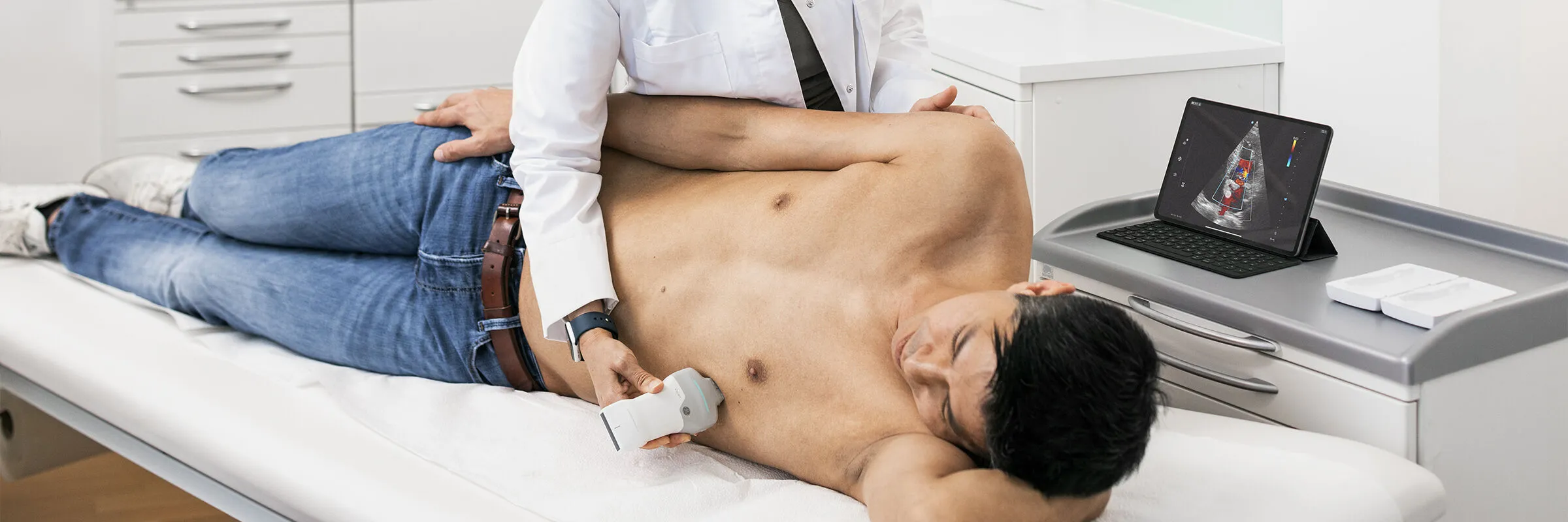
The Vscan Air CL is specifically aimed at point-of-care applications in orthopedics, rheumatology, sports medicine, and general medicine.
Particularly relevant features of the device for MSK medicine are:
- Wireless design
- Dual-probe system without probe changes
- Linear probe for tendons and ligaments
- Curved-array probe for deeper regions
- Mobile use in practice, outpatient clinic, or acute care
This combination of mobility and integrated probe architecture proves particularly advantageous in everyday clinical practice, as examinations can be carried out without changing devices and independently of a fixed examination location.
Dr Sebastian Schellhaas, a GP and sports medicine physician in Groß-Biberau, describes how much of a difference this makes in everyday practice: “The device is small and handy. I can put it in my jacket and move effortlessly from one patient to the next – and on top of that, the image quality is also far superior.”
Compared with classical console systems, the wireless design substantially facilitates dynamic examinations. At the same time, the elimination of separate probes reduces interruptions in the examination workflow. Particularly for time-critical examinations or frequently changing settings, this can significantly simplify the workflow in practice and hospital.
Further information on use in musculoskeletal diagnostics can be found on the page about the mobile musculoskeletal ultrasound device. If you would like to take a closer look at the differences between the two Vscan models, the comparison can help.
Frequently Asked Questions about MSK Medicine
Which Specialties Perform MSK Ultrasound?
MSK ultrasound is used primarily in orthopedics, rheumatology, sports medicine, trauma surgery, and general medicine. Musculoskeletal sonography is also increasingly part of everyday clinical practice in physical medicine and rehabilitation.
Which Probe Is Suitable for MSK Examinations?
For superficial structures such as tendons, ligaments, or small joints, high-frequency linear probes between approximately 5 and 15 MHz are suitable. Deeper regions such as the hip benefit from curved-array probes with greater penetration depth.
Does MSK Ultrasound Replace MRI?
Not entirely. MSK ultrasound is excellently suited to many superficial and dynamic questions and provides rapid examination directly at the point of care. For complex intra-articular or deep structures, however, MRI often remains the complementary or further imaging modality.
JB09710xe
Other Topics

How handheld ultrasound can impact practice in Primary Care – featuring GE HealthCare's Vscan Air™ family with Vscan Air™ CL and Vscan Air™ SL

Lung Ultrasound: Recognizing Findings and Making Diagnoses

Mobile Ultrasound in Emergency Medical Services: Use, Benefits, and Selection
Ejection Fraction: Significance, Normal Values, and Measurement

Bladder Volume Measurement by Ultrasound: Methods, Formula, and Application

Empowering nurses through innovation: Vscan Air™ handheld ultrasound in action at the ICN Congress 2025

A better outlook for mothers in Ethiopia: How Vscan Air™ is transforming maternal care in remote regions

The benefits of handheld ultrasound with AI-based Auto Bladder Volume measurement in everyday clinical practice

Experiences with handheld ultrasound in obstetrics

Vscan Air™ CL ultrasound combined with Usono ProbeFix Dynamic: Observation of muscle activity in real-time



 Belgium
  Brasil
  Bulgaria
  Croatia
  Czechia
  Denmark
  Deutschland
  España
  Estonia
  Finland
  France
  Greece
  Hungary
  Ireland
  Italia
  Latvia
  Lithuania
  Luxembourg
  Malaysia
  Netherlands
  Norway
  Österreich
  Poland
  Portugal
  Romania
  Slovakia
  Slovenia
  Sweden
  Schweiz
  UK
UK 
